Using AI to help diagnose brain aneurysms
Date:
Changed on 02/04/2025

René Anxionnat: It is estimated that 3% of French people currently have aneurysms, which are like hernias affecting the blood vessels within the brain, primarily at branch points. If these aneurysms rupture this causes bleeding, the consequences of which can be extremely serious. Prior to the 1990s the only treatment option was surgery, before the development of an endovascular treatment involving sealing off the aneurysm from the inside using coils. This remains the most common treatment option, although other methods including stents and flow diverters have also been devised. This falls within the bracket of interventional neuroradiology, a mini-invasive medical discipline developed in the 1960s. Among its pioneers was Professor Picard, who founded the Department of Neuroradiology at Nancy Regional University Hospital. Since then it has continued to evolve from a clinical, technical and scientific perspective. A risk-benefit analysis is carried out for each individual case, meaning that not all aneurysms that are discovered are treated. Magnetic resonance imaging (MRI) has improved the detection of unruptured aneurysms and plays a vital role when it comes to deciding on treatment and monitoring.
Erwan Kerrien: All treatments involve the use of x-ray imaging, which significantly reduces the risk of intraoperative trauma. However, these images are 2D, whereas the vascular structure that interventional neuroradiology is concerned with is extremely complex and 3D. 3D imaging does exist, whether in the form of computed tomography (CT) scans or MRI, but interpreting these images and linking them to those obtained during procedures is a mammoth undertaking. This is why the involvement of specialists in image processing is important, like us, researchers from Tangram, whose work concerns image analysis and realistic physical and geometric modelling.
The goal is to develop a simple, reliable tool for automatic detection
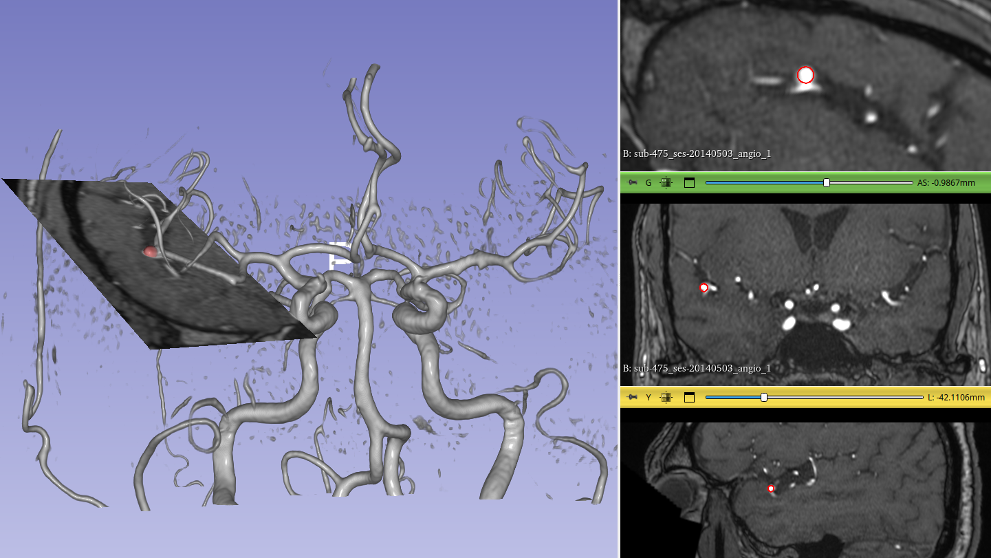
Erwan Kerrien: The challenge lies in the fact that aneurysms are very small - the average diameter is between 5 and 8 mm - and can be found in multiple locations. To detect them, not only do you need images of sufficient quality, but you also need the capacity to interpret them. This will depend on the level of expertise of the radiologist, the equipment they have at their disposal for analysis and how tired they are. With this in mind, the aim was to develop a tool that uses artificial intelligence to help practitioners make diagnoses by automatically detecting aneurysms. The algorithm is centred around a deep neural network architecture developed by Youssef Assis, a PhD student we began supervising in 2020 and who completed his PhD thesis in 2024, jointly funded by Nancy Regional University Hospital and the Grand Est region as part of the French National Research Agency (ANR)’s PhD funding programme. The next step was to validate the effectiveness of the tool. This was the responsibility of Liang Liao, another PhD student who is also a hospital practitioner and who joined the project in 2021. Our aim was to evaluate the performance of experts in comparison to non-experts and to determine how AI, through our algorithm, could improve the latter. In our initial results non-experts had an average detection rate of 75%, meaning they detected 3 out of 4 aneurysms. The detection rate for experts, meanwhile, was between 84 and 90%, depending on whether their analysis was cursory or more detailed. The rate with automatic detection was 86%. What this shows is that, in all cases, AI improves detection.
René Anxionnat: The goal is to provide doctors with a simple, reliable tool for automatic detection and to help them detect aneurysms, irrespective of their size. But AI isn't going to replace the doctor-patient relationship, and decisions on treatment will continue to be made by doctors.
Technological breakthroughs made will be used for everything relating to medical imaging.
René Anxionnat: The aim is to target practical applications that will help to improve patient care. This might involve incorporating tools into MRIs for the automatic detection of aneurysms or treatment decision support based on morphological factors such as an individual’s height or body shape. Technological feasibility is one thing, but doctors also need to be convinced of the usefulness of such tools. In the early days of 3D angiography, some felt it would only be of use in very specific cases, and yet it is now used routinely to treat all brain aneurysms.
Erwan Kerrien: The models we have developed - which are free to access online - have still to undergo clinical validation. The program is trained using a base of 270 volunteers, which isn't enough to certify our results. Training the tool using much larger databases will enable us to improve its performance. Nancy Regional University Hospital is involved in the creation of a national register coordinated by the French Society of Neuroradiology (Société française de neuroradiologie, SFNR), compiling 5,000 data points on patients which will then be expertly annotated. Technological breakthroughs made in the context of the automatic detection of aneurysms will be used for everything relating to medical imaging, making it possible to extend the use of minimally invase techniques to other medical conditions.
Erwan Kerrien: I joined the Department of Interventional Neuroradiology in 1996 as part of a Cifre PhD (industrial agreements for training through research) that I was doing at General Electric, specialists in medical imaging, in collaboration with Inria. I spent a lot of time with René Anxionnat within the department, learning about what working conditions were like for staff there and the challenges they had to deal with. My background in engineering had seen me work with mathematics and computer science, and the ideas I was coming up with weren’t necessarily of interest to doctors, as interesting as they might have been from a theoretical point of view. The first step involved spending time in operating theatres, speaking to doctors and observing cases so as to be able to identify needs. Aneurysms emerged as a subject in 2019 when developments in deep learning made it possible to process bigger volumes of data.
René Anxionnat: I followed a similar route, studying for a PhD in Computer Science with the Loria laboratory. This has given us an opportunity to get to know each other, to figure out how we both might contribute and, over time, to identify practical subjects for us to work on. We each came along with our own expectations and needs and we are moving forward together, striving to ensure that we learn from and complement one another. A working relationship spanning 30 years has to be built on trust, but that doesn't happen automatically - it’s something you need to work on.
Erwan Kerrien: Trust, yes, and also humility. You have to be able to look beyond your own convictions and preconceptions. This has been a hugely rewarding experience on both a scientific and a human level.
A graduate of Télécom Paris, Erwan Kerrien completed his PhD in Computer Science in 2000 at the Institut Polytechnique de Lorraine – in partnership with GE Medical Systems (now GE Healthcare), Inria and the Department of Interventional Neuroradiology at Nancy Regional University Hospital – before joining Inria the following year as a researcher assigned to the Loria laboratory. He is currently a member of the Inria project team Tangram.
After completing his PhD in Medicine in 1989, René Anxionnat joined the Department of Clinical Neuroradiology at Nancy Regional University Hospital. In 2003 he completed a PhD in Computer Science focusing on methods and tools for the removal of arteriovenous malformations in the brain in a multimodal context. He is now a university lecturer and hospital practitioner in diagnostic and interventional neuroradiology at the University of Lorraine, in addition to his role as head of the Department of Interventional Neuroradiology at Nancy Regional University Hospital.