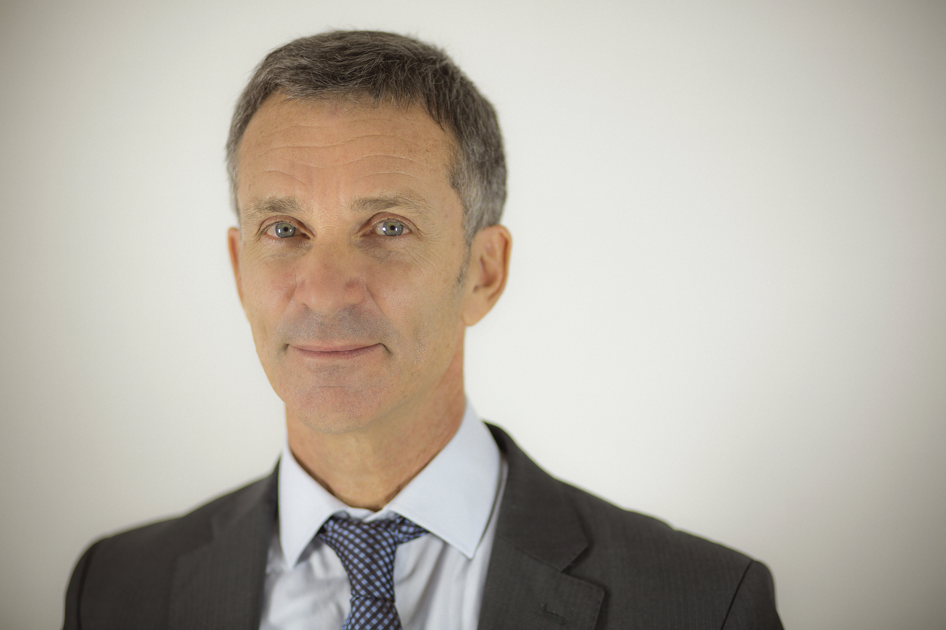
Nicholas Ayache: "Medical Imagery and I.T.: the personalised digital patient"
Date:
Changed on 24/02/2021

Medical imagery has made exceptional progress over the last 30 years. Today it enables the structural and functional properties of tissues and organs to be captured on various scales: macroscopic for organs, microscopic for cells and even nanoscopic on a molecular scale. Current research into imagery aims to assist the clinician in the analysis of this ever-increasing volume of information by integrating the entirety of this data into multi-scale, multimodal 3D images (obtained through a variety of techniques).
The ERC MedYMA project intends to go one step further by integrating the temporal dimension too , in order to take into consideration the dynamic properties of an organ e.g. cardiac movement, the dynamics of a disease, and indeed the growth of a cancerous tumour or the atrophy of cerebral regions due to Alzheimer's disease. In the first example, the objective is to be able to determine as soon and as accurately as possible whether a movement anomaly exists. The two remaining examples involve quantifying the progression of the disease observed between two examinations. It also enables medical staff to determine more swiftly the effectiveness of a treatment in order to change it swiftly if necessary, or indeed to make use of the simulation to estimate, in advance, the most suitable treatment for the patient's disease.
To incorporate all of this data, we suggest adjusting biophysical models in order to make them specific to each patient. These generic models are thus personalised thanks to the medical images. They are constructed from the physical and biological properties of the organs or tissues, taking account of the statistical variability existing between individuals. One original aspect of the ERC MedYMA is the construction of computer-generated medical images based on these biophysical models. These computer-generated images will make it possible to validate the analysis algorithms developed during the project, but also to design new analysis algorithms based on modern I.T. learning methods. These computer-generated images will therefore need to be as realistic as possible: this is one of the major scientific challenges of this project!
The computer analysis of the medical images, by exploiting biophysical and statistical models of living beings, enables better interpretation of the patient's medical examinations, simulation of the development of a disease and the effectiveness of a therapy to be predicted.
In partnership with the Inria Sisyphe, Macs and Reo teams, the Asclepios team has already developed an initial biophysical model of the heart, based on the organ's geometry and on its electrophysiological and mechanical properties. The ERC MedYMA project will make it possible to improve this personalised virtual heart model, in order to help quantify normal movement and detect anomalies (arrhythmia, heart failure, etc.). It may also be used to simulate a therapy (radiofrequency ablation, fitting of a pace-maker, etc.) and to predict the expected benefits for the patient. Indeed, today around 30% of patients fitted do not receive the real benefits of their pace-maker. In oncology - another of our fields of application together with neurology and cardiology - it is hoped that the models will enable the target of the radiotherapy or surgery to be refined by taking better account of the non-visible infiltration of the tumour by the simulation.
The ERC grant makes it possible to plan over a longer period than usual, and therefore to conduct more fundamental research , with very few administrative constraints. It will mainly finance PhD students, as well as several post-doctoral researchers and engineers as, above all, it involves research into algorithmics and applied mathematics, with a small amount of software engineering. This work will be conducted within the Asclépios team, whose expertise - the researchers Hervé Delingette, Xavier Pennec and Maxime Sermesant are involved in the project - will contribute to the success of the undertaking. The grant will also facilitate work with our academic and clinical partners in France as well as in Europe and the United States, particularly with France's three new university health institutes (IHU), with which we collaborate for modelling of the heart (Bordeaux), the brain (Pitié-Salpêtrière in Paris) and the digestive system (Strasbourg). Industrial partners will also be involved throughout the project. This is an indispensable element in order to ensure effective transfer of innovations.
The creation of a generic patient model within MedYMA should constitute a break in the capacities for automatic interpretation of medical images and in clinical practices. These models, built based on the biophysical properties of organs and knowledge of pathologies, enable the creation of virtual hearts, livers and hearts, and the simulation of the occurrence and development of pathologies such as tumours, arrhythmias, atrophies, etc.
These computer-generated images may be generated at will and in very large quantities in order to establish larger, more varied databases than the patient database that are most often incomplete and difficult to access. For example, it is possible to grow tumours by modifying the proliferation and infiltration parameters in order to obtain a very extensive range of intermediate cases.
Such databases are of interest in order to test the reliability of image-analysis and diagnosis-assistance software. They are also of interest in leading the software designed in order to refine their detection capacity through learning on numerous cases. Another application envisaged consists of generating computer images covering the largest possible number of cases in order to coordinate a medical version of a flight simulator for pilots. Practitioners may in this way be confronted, during the course of their training, with extreme, highly-varied or extremely rare situations, providing them with the most extensive expertise possible.