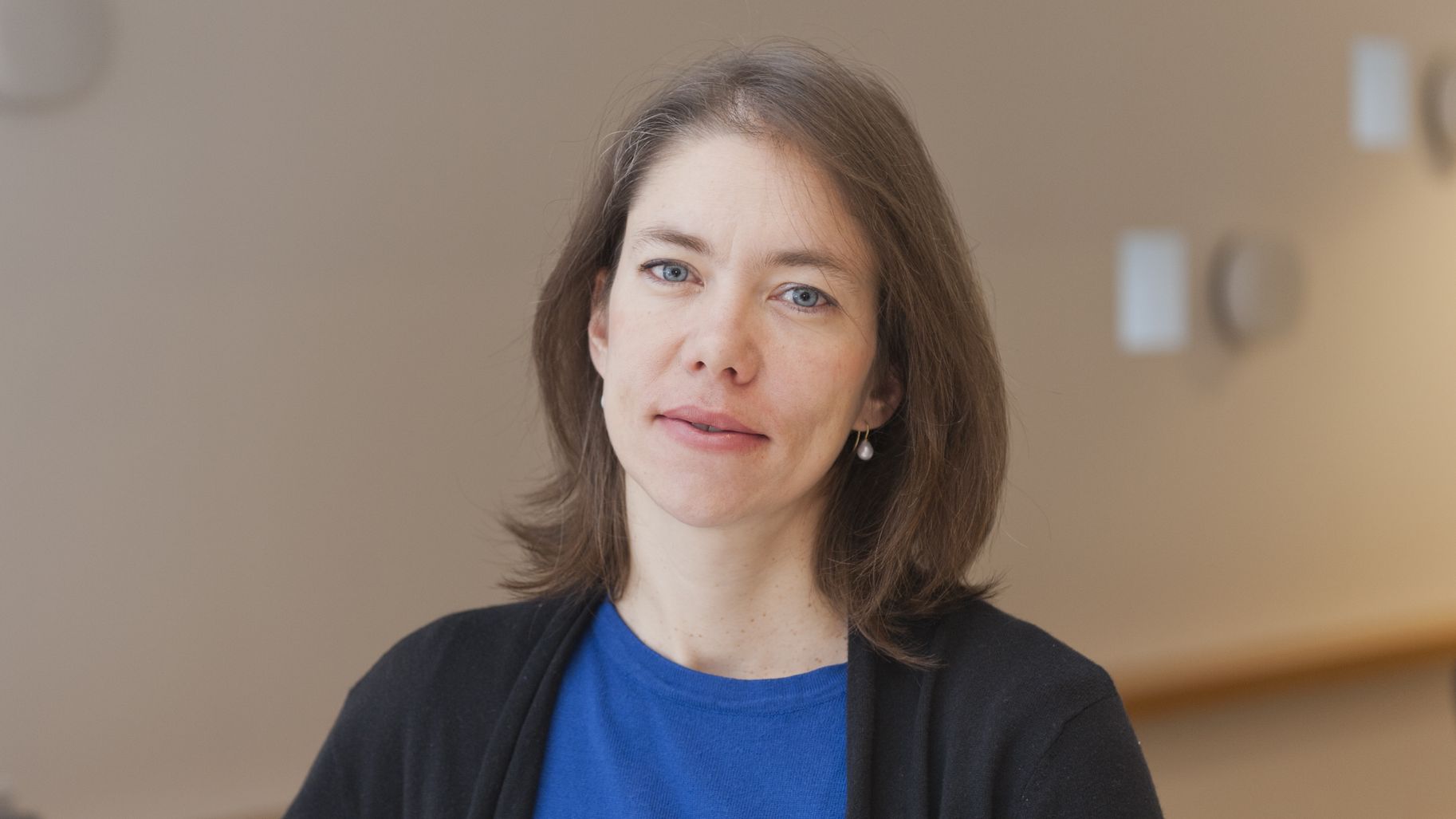
Irène Vignon-Clementel - digital simulation to help surgeons
Date:
Changed on 06/05/2020

It was while studying for her master’s degree in applied mathematics and theoretical physics at the University of Cambridge that Irène Vignon-Clementel took a course taught by Professor Pedley on biofluids. This experience prompted her to apply fluid mechanics to the field of health.
He (Professor Pedley) really sparked my interest in this type of work. I then studied for a PhD at Stanford University in the USA with Professor Taylor, which I found extremely interesting. I wanted to push further ahead with clinical applications for digital simulations”, she explains. “I wanted to help people and society as a whole by bringing together science and medicine.
The Cardiovascular Biomedical Research Laboratory at the University of Stanford, where the young researcher studied for her PhD between 2001 and 2006, was one of the first in the world to use patient data in digital simulations. Upon returning to France, Irène Vignon-Clementel was immediately given a position as a researcher at Inria as part of the REO team, where she began working on a project aiming to study the different parts of the heart.
"One of the reasons I was chosen was probably my experience in using patient data, which I had acquired at Stanford University. At the time, this sort of research didn’t exist in France. What attracted me to Inria was how open the institute is to interdisciplinarity and its desire to push for transfer, whether it’s to companies or hospitals, as well as really supporting researchers."
Irène Vignon-Clementel began working on different congenital heart diseases, including deformities of the heart or associated vessels. Such diseases are quite complicated for patients, and she had relatively little data at her disposal.
I also found it interesting getting to work with doctors, trying to understand their problems and helping them to tackle challenges and to find answers to their questions, she explains.
Over the next few years, Irène worked on ventilation, respiration and deposits from aerosols (particles) in the lungs. At the same time, she also began research on the liver. “In 2012, I was contacted by Professor Vibert (APHP*, Inserm**), who was looking to develop a better understanding of post-op issues affecting the liver, an organ which is capable of regenerating after partial removal. The goal was to find out whether not it would be possible to use digital simulation in order to predict changes in blood pressure and blood flow after surgery”, she explains.
Irène Vignon-Clementel and her colleagues from Paul Brousse hospital in Villejuif are currently working with Professor Line from Oslo University Hospital, a Norwegian surgeon who developed the RAPID technique: a method which involves performing a gradual liver transplant over different stages so that only part of the grafts are used. This technique could prove highly efficient when it comes to performing transplants on sick patients while “economising” the available organs. However, as things currently stand, it can lead to high blood pressure at certain points during the operation. Using a digital simulator would make it possible to better understand how this hypertension is produced, to identify cases where the RAPID method could be used and to optimise the use of grafts for patients.
The ERC Consolidator Grant will enable Irène to pursue her research into the liver and congenital diseases. Her team is now capable of explaining and modelling changes to blood flow and blood pressure in animals’ livers following removal. The team’s aim is now to model different aspects of blood flow in humans.
“We will be focusing on whole organs, but also on different parts of them and at different levels, including at a microscopic level”, Irène continues.
We want to know the effect certain conditions have on the organs, such as pulmonary hypertension or cirrhosis, for example. We will then attempt to employ data from non-invasive imaging, obtained using either MRI or scanners. If we get better at using this data, this will enable us to develop a better understanding of the condition of organs prior to surgery.
The team’s long-term goal is to design a digital simulator that will make it possible to plan medical operations by taking the condition of patients’ organs into account, and which will be used in practice by clinicians.
“This ERC Consolidator Grant is perfect for our project, given the various different parts we will be studying and the fact that we will need to call upon a number of different specialists”, stresses Irène. This funding will enable us to recruit young clinical researchers, as well as young researchers in simulation. Ordinarily, these types of funding are separate. This grant is staggered over a period of five years, and we are hopeful that we can design the simulator during this time.”
Her hopes for the next few years?
I would like to improve the way in which illnesses and surgery are understood in order to provide surgeons and cardiologists with solutions that will enable them to better anticipate their acts and surgery-related issues.
Digital simulation can prove highly useful for doctors with little experience with such conditions, as well as for training purposes. This type of tool can also be used to test ideas for new operations, limiting the risk for patients and better anticipating how their organs will respond to surgery.
*APHP: Assistance publique hôpitaux de Paris (the university hospital trust operating in Paris and the surrounding area)
Inserm: Institut national de la santé et de la recherche médicale - The French National Institute for Health and Medical Research
The project is funded by the European Research Council (ERC) under the Horizon 2020 program.