Prostate cancer: digital sciences to assist diagnosis
Date:
Changed on 08/01/2025

The prostate is a gland in the male genital tract located between the bladder and the rectum. From the size of a chestnut until the age of fifty, its volume naturally increases with age, along with the risk of cancer, which is rare in younger people. The french National Cancer Institute (INCa) considers the prognosis for this disease to be ‘good, even very good’. Even so, it is the third leading cause of cancer death in men. In 2021, it was the cause of 9,200 deaths.
However, the mortality rate is falling: -2.4% per year on average between 2011 and 2021, according to INCa, which explains this progress by improved treatments and access to screening. In fact, 80% of prostate cancers are detected when they are still localised. Diagnosing the disease early is therefore vital.
Screening begins with the measurement of a specific antigen, PSA (Prostate Specific Antigen). But this is not a sufficient indicator. It therefore leads to additional tests: a rectal examination and, if necessary, a biopsy. This procedure involves taking tissue samples from different areas of the prostate with a needle. It can be painful and, like any operation, can lead to complications. The most serious complications, such as infections or acute urine retention, account for less than 5% of cases.
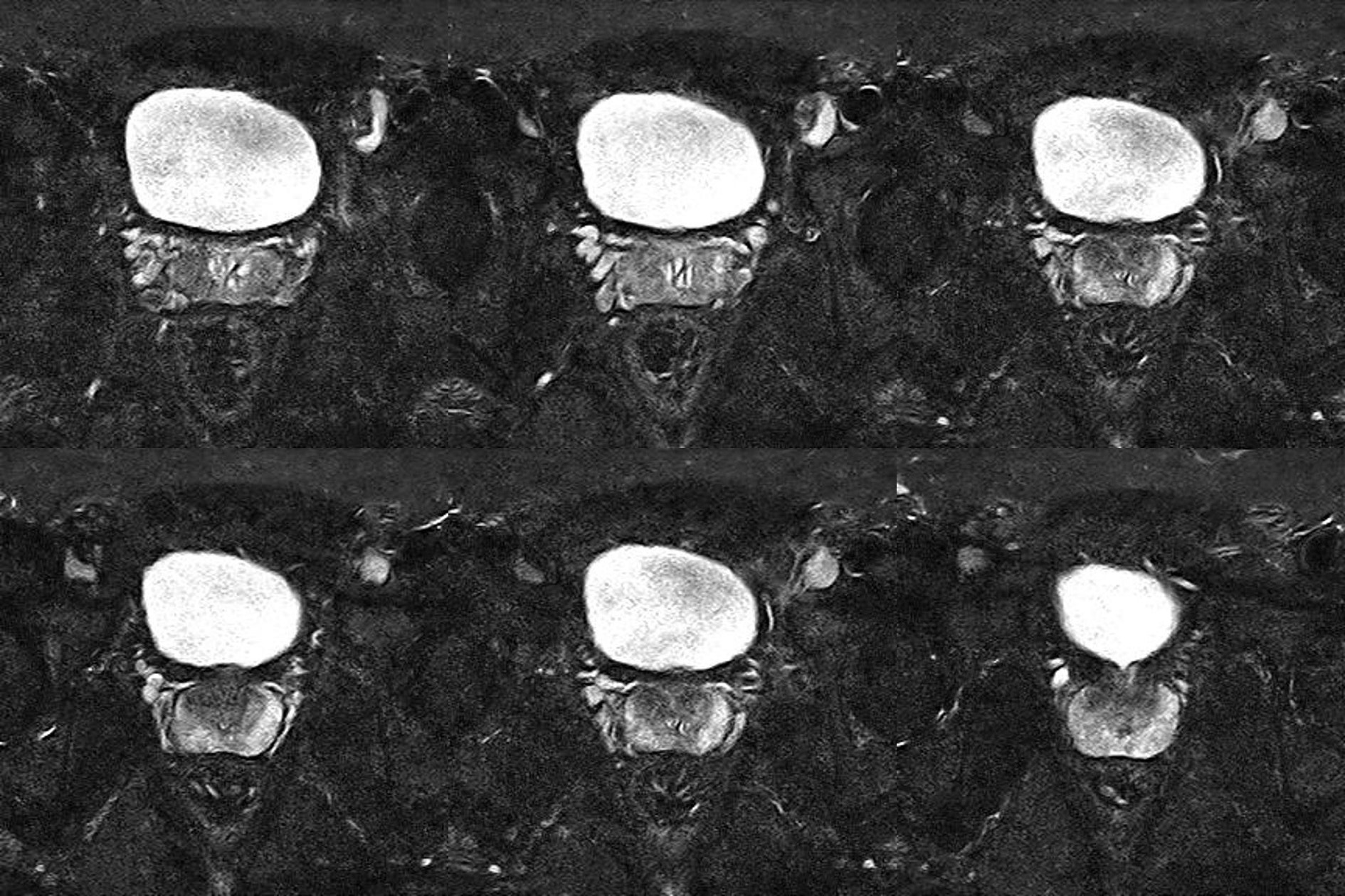
However, prostate lesions are common, not always significant, and the disease progresses slowly. As a result, screening is not routinely recommended in France, and biopsies are best reserved for the most suspicious cases. But how do you identify them? ‘Magnetic resonance imaging (MRI) can help,’ says Jan Ramon, Research Director at the Inria Centre at the University of Lille, and a member of the Magnet project team, working with the CRIStAL laboratory (CNRS, university of Lille, Centrale Lille).
This is one of the objectives of the Flute European research project, which since 2023 has brought together twelve academic and industrial partners. ‘One of them had a model for determining the need for a biopsy for a given patient, based on seven variables, including age’, explains the researcher. ‘But this could be improved with more data. We are therefore looking to build a more robust model, based on image recognition, which is also capable of locating the areas where the samples should be taken.’
To achieve this, a number of difficulties need to be overcome. Firstly, the machine learning approach requires a large amount of training data: more than a single hospital could provide. Secondly, certain statistical variations between populations could create biases. An algorithm trained on data from Spanish patients would not necessarily perform in the same way on a Belgian population, for example. Data sources must therefore be sufficiently diversified. Hence the importance of international collaboration!
‘We work with three hospitals, in Spain, Italy and Belgium’, explains Jan Ramon. ‘Based on this geographical distribution, the aim is to obtain a generic model that can be applied everywhere, requiring only adjustment to local specificities on the basis of a small sample of data’. However, hospitals cannot share confidential health data, and even less so abroad.
The development of the image recognition model begins with training on different organs: prostates, lungs, kidneys, etc. ‘The aim is to learn what medical images look like’, comments the researcher. Specialisation then begins with raw MRI images of prostates, with no annotations. This stage can be completed with synthetic data. These do not correspond to any real patient, but have similar properties and can be generated in large quantities.
Image

Verbatim
The third training phase uses real MRI data annotated by specialist radiologists to teach the model to differentiate between a healthy and diseased prostate. This data is more expensive to produce, and only available in smaller quantities.
Auteur
Poste
Research Director at the Inria Centre at the University of Lille
To share this real data, Flute relies on a platform developed as part of another international project, Trumpet. It uses federated learning: a method that enables learning algorithms to be trained locally, before the resulting models are pooled. ‘This system guarantees that no confidential data, which could identify patients, can be extracted from these models’, emphasises Jan Ramon. ‘But this platform is not suitable for exchanging large volumes of data, such as that generated by medical imaging.’
By April 2026, Flute aims to develop two tools: an algorithm for diagnosing prostate cancer using MRI data, and the adaptation of the Trumpet platform to these large volumes of data. In the longer term, these tools could be applied to the diagnosis of other pathologies.
Progress in image analysis should therefore help to improve diagnosis, but is not intended to replace biopsy. Biopsies are still necessary for a more accurate assessment of the seriousness of a case of cancer. But here too, digital science research could improve patient care. This time, the focus is on modelling and robotics.
Biopsies could be automated to improve precision. Whether performed transrectally or transperineally, biopsies need to aim precisely at the targeted areas - while avoiding the urethra, which passes through the prostate. To achieve this, an automated system needs to be guided by real-time imaging: ultrasound or MRI. This involves several challenges: guiding the medical robot and modelling the prostate in its environment.
The Inria Defrost project team, working jointly with the CRIStAL laboratory and Centrale Lille, is looking at deformations in robotics. ‘We worked on a robot carrying a flexible cylinder, on which the ultrasound probe and needle are mounted’, explains Yinoussa Adagolodjo, professor at the university of Lille and member of Defrost.
The deformations of this flexible element, designed to cushion contact with the patient, must therefore be taken into account when controlling the robot and positioning the probe. ‘To model them, we used specific mathematical models, different from those used in conventional robotics’, stresses the researcher. Next comes the modelling of the inside of the body and its deformations.
Image

Verbatim
We start by using MRI images to map the prostate and surrounding organs in the pelvic region, which is a complex area. There are many connections and interactions between organs and bones, and these are difficult to model accurately. We therefore use a simplified model. This model is then parameterised using data from the scientific literature about the mechanical properties of the organs, such as their rigidity.
Auteur
Poste
Professor at the university of Lille, member of Defrost project team
The algorithms developed enable the needle to be aligned with the target. The needle is then injected at high speed to minimise tissue deformation. The team modelled the entire operation, demonstrating its feasibility in a number of scenarios worked out with a doctor.
As well as designing medical robots, this work is being used to make ‘phantoms’: replicas of organs that reproduce their characteristics and behaviour, for training or experimental purposes. ‘During a biopsy, repeated punctures cause the prostate to swell’, explains Yinoussa Adagolodjo. An initial phantom designed by the team reproduces this behaviour, and a thesis in the Defrost team is currently continuing this work.
‘The aim of this thesis, carried out by Sizhe Tian, is to create an active phantom capable of reproducing variations in prostate volume and rigidity, whether due to ageing or carcinoma’, explains the researcher. ‘This tool could be used to train doctors in rectal examination, or to test the algorithms of our robots. In the longer term, with the modelling work carried out in this context, we can even imagine automating and standardising this procedure, the outcome of which is still very much dependent on the practitioner’.
In the future, the medical community should be able to draw on new tools from research in machine learning, modelling and robotics to improve prostate cancer diagnosis. As things stand, however, the screening remains limited to specific cases, because of ‘its disadvantages and the uncertainties about the benefits’, writes INCa, which stresses the importance of information and recommends above all that those concerned discuss the matter with their doctor.
- Prostate cancer screening in question (in French), Institut national du cancer.
- Prostate cancer : forewarned is forearmed (in French), France Culture, 15/01/2024.
- Prostate cancer: transgender women remain at risk (in French), Pourquoi docteur, 04/05/2023.
- Medical imaging: can artificial intelligence deliver?, Inria, 11/05/2022.
- Magnet prescribes federated learning to healthcare facilities, Inria, 9/5/2022.
- Organs, robots: simulating deformations (video, in French), Inria, 06/11/2024.